English
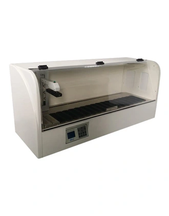
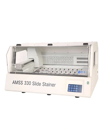
The AMOS Slide Stainer is a highly advanced instrument designed for automated staining of histology slides. The automatic staining machine offers precise and efficient staining protocols, ensuring consistent and reproducible results. As a premier automated slide stainer histology solution, it integrates high-capacity processing with advanced fluidics. The stainer accommodates multiple slides and provides a user-friendly interface for easy operation. The AMOS Slide Stainer is available for sale, providing a reliable and efficient solution for slide staining in research, diagnostics, and educational laboratories.
1. Routine Hematoxylin and Eosin (H&E) Staining: H&E staining is the most commonly performed staining technique in histology and pathology. It involves the use of hematoxylin to stain cell nuclei blue-purple and eosin to stain cytoplasm pink. Slide stainers automate the process by precisely timing and controlling the staining steps, ensuring consistent and reliable results for the visualization of tissue structures. These systems are widely referred to as an automated H&E stainer in modern laboratories.
2. Immunohistochemistry (IHC): Slide stainers are widely used for immunohistochemistry, a technique that allows the detection and visualization of specific proteins in tissue samples. The slide stainer automates the incubation steps with primary antibodies, secondary antibodies, and the chromogen, providing precise timing and temperature control. This ensures reproducibility and accurate protein detection in diagnostic and research applications.
3. Special Stains: Slide stainers are capable of performing a variety of special stains that allow visualization of specific tissue components or abnormalities. These systems, also known as automated histology stainer, support stains such as silver staining, Masson's trichrome, and PAS staining. Modern laboratories often rely on a staining machine for histology to ensure standardized protocols and reproducible results.
4. Fluorescence In Situ Hybridization (FISH): Slide stainers equipped with fluorescence capabilities can perform FISH staining. This technique utilizes fluorescently labeled DNA probes to target specific DNA sequences in the tissue sections, allowing for the detection of genetic aberrations, such as gene amplifications or chromosomal rearrangements. Slide stainers, as an automated staining machine, automate the various hybridization and wash steps involved in FISH, ensuring reproducibility and accuracy in the detection of genetic changes. Advanced systems such as a fully automatic slide stainer or fully automated slide staining instrument are commonly used in modern histopathology laboratories.
These staining techniques, performed with the assistance of automatic slide stainer histology systems and histology autostainer platforms, are essential for visualizing tissue components and identifying pathological changes. A modern tissue stainer or automated histology stainer significantly improves efficiency and reproducibility in histopathological analysis. Many research labs also prefer a slide stainer for research to support experimental workflows.
As a versatile histology autostainer, it supports multiple staining protocols and improves workflow efficiency in histopathology labs. The implementation of an automatic slide stainer in histopathology ensures that every rack of slides receives identical treatment.
1. Automation: Slide stainers automate the staining process, reducing the reliance on manual labor and minimizing the risk of human error. Automated protocols ensure consistent timing, temperature control, and reagent dispensing, leading to reproducible and reliable staining results. These systems are also referred to as automatic staining machine in histopathology in modern laboratories.
2. Time savings: The automatic stainer in histopathology significantly reduce the hands-on time required for slide staining. By streamlining the staining process, slide stainers allow laboratory staff to focus on other tasks, increasing overall laboratory productivity. Batch processing capabilities enable multiple slides to be stained simultaneously, further saving time. Many laboratories compare different models of staining machine for histology when selecting equipment based on workflow needs.
3. Staining consistency: Slide stainers, as automatic stainers in histopathology, ensure consistent and standardized staining protocols, eliminating variations that may occur with manual staining methods. Consistency in staining improves intra-laboratory reproducibility and allows for accurate comparison of slides over time or between different laboratories. This is a key feature of autostainer histology systems and automated stainer histopathology platforms.
4. Reagent efficiency: Slide stainers optimize reagent usage, reducing waste and cost. Automated dispensing of reagents minimizes overconsumption and contamination risks, ensuring that reagents are used efficiently without compromising staining quality. This is particularly important for expensive or limited quantity reagents utilized in specialized stainings. Procurement decisions often consider staining machine price and overall operational cost of an automatic slide stainer and hier machine in histopathology laboratories.
5. Workflow integration: Slide stainers can be integrated with laboratory information management systems (LIMS) or digital imaging systems, streamlining the workflow and improving data capture and traceability. Connected systems enable efficient tracking of slides, reagents, and staining protocols, promoting seamless integration with downstream processes for analysis and archiving. For laboratory procurement planning, factors such as slide stainer price and system configuration are often considered when selecting an appropriate model.
E-mail:
Tel:
Whatsapp:
Address:
24, 632-642 Clayton Road, Clayton South VIC 3169, Melbourne, Australia